| Dec 2022 Issue 20 | ||||||
 |
||||||
|
|
| Research | ||
| For tofu to tissue-sectioning: the cutting-edge imaging solution transforming the research world | ||
|
|
||
|
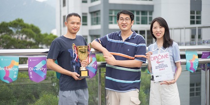
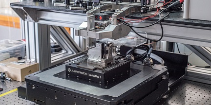
When Professor Chen Shih-chi from the Department of Mechanical and Automation Engineering and his team began developing a new precision tissue-cutting machine for pathological research seven years ago, one of the first things they experimented with was tofu. Today, this humble staple of Asian cuisine can be credited for transforming the face of 3D imaging and drug screening, as the team’s multi-patented novel tissue-sectioning solution continues to make waves in top research communities across the world. A microtome is an instrument that sections biological specimens into very thin slices for microscopic examination; however, traditional microtomes have only been able to process fixed or hardened tissues. To enable the sectioning of soft or live tissue into slices of desired thickness for whole brain/organ super-resolution imaging, novel solutions were necessary – which is why Professor Chen’s precision engineering expertise was sought by leading research groups to explore new ways that could open a new chapter in brain research, such as the construction of 3D neural connection maps. The microtome can also minimise cellular damage while maintaining high cell viability, even on sectioned surfaces. As a result, the precision-sectioned slices can be cultured in vitro for a long time, allowing for drug screenings and other new applications. Furthermore, the team has gone a step further by combining the microtome’s automatic 3D tissue sectioning system with a fluorescence microscopy imaging platform to realise the first-ever low-cost super-resolution 3D imaging solution. The solution’s effectiveness was proven through the 3D super-resolution imaging of single neuron dendritic branches in a mouse’s brain (<100nm) during the development process. Ultimately, through the continual pushing of boundaries, Professor Chen and his team’s novel solution has successfully opened up new possibilities for more exciting breakthroughs to come in research and beyond – promising a better world for all. More details: Enterprising startups star at CUHK’s Entrepreneur Day | CUHK in Focus |
||
|
||
 |
||
|
|
|||||||||||||||||||||||


