| Jan 2018 Issue 6 | ||||||
 |
||||||
|
|
| Research | |||||||||
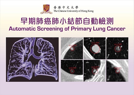
| Automated Medical Image Analysis Using Deep Convolutional Neural Networks | |||||||||
|
Prof. HENG Pheng Ann, Department of Computer Science and Engineering, CUHK With the large number of new cancer cases registered and the public's raised awareness of health, it is not surprising to find a rapidly growing demand for improved services in the medical sector. Medical image analysis has been playing a crucial role in the modern healthcare industry. The artificial intelligence platforms (AI) we developed are able to provide a reliable, scalable as well as cost-effective alternative to significantly reduce the tedious workload of the doctors and greatly improve the diagnostic accuracy [1].
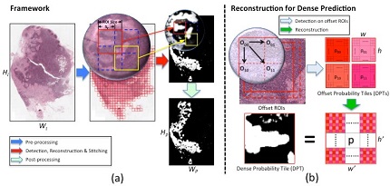
Detection of pulmonary nodules through Deep Learning We developed a two-stage framework to detect the lung nodules from CT images [3]. Firstly, we designed a 3D neural network that incorporates the particular structural characteristics of thoracic CT images and is able to predict suspicious nodule locations by finding high-probability regions in the 3D score-map. Secondly, we conduct false positive reduction that aims to remove those candidates that are not true nodules. This task is the most difficult and challenging component in the detection system, as many of the false positives take quite a similar appearance to the real nodules. Our AI platform can locate the pulmonary nodules from CT images within 30 seconds, with a sensitivity of over 91%. Automated Detection of Metastatic Breast Cancer in Histology Images To solve the problem, our team has developed a novel deep cascaded convolutional neural network to process the histopathological images. Making use of a fully convolutional network, the model can efficiently and accurately detect metastatic cancer with a high-resolution score-map. The whole automated analysis process takes about 5~10 minutes, as compared to the 15~30 minutes examined by the naked eye. In terms of accuracy, the system has achieved a rate of about 99 percent, which is 2 percent higher than analysis conducted by experienced pathologists. This indicates that it is an invaluable reference for clinical diagnosis of breast cancer. The projects take full advantage of our research experience in the past few years, a series of research results and new technologies/methods obtained, including medical image processing and deep learning techniques. In addition, frequent collaborations among the AI researchers, IT experts, and the medical professionals involved in this project, has greatly helped to develop the platforms and improve both the efficiency and accuracy in diagnosing the diseases as well as the treatment provided to patients in the healthcare system. In general, our project provides a well-established AI platform for computer-aided cancer diagnosis and monitoring in the medical community of Hong Kong, with global potential. Public Hospitals and Imaging Centres can adopt the platform to support the limited number of doctors and help them stabilise their performance by reducing significantly the misdiagnosis rate, which is due to limited examination time. Currently, we have established collaborations with top hospitals in HK and Mainland China. Our developed products have been set up in many hospitals including West China Hospital and United Family Healthcare. References
|
|
||||||||
|
|
|||||||||
 |
|||||||||
|
|
|||||||||||||||||||||||